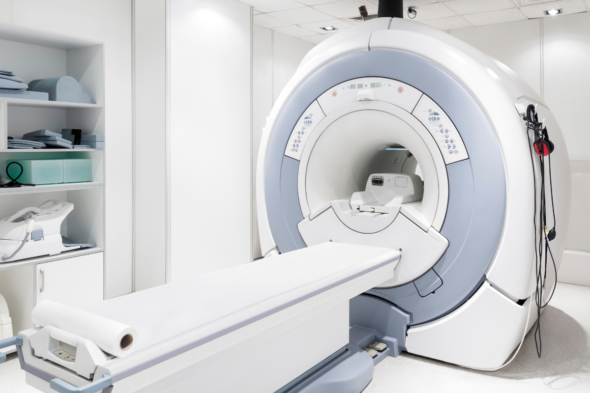
Doctors can diagnose a number of conditions with imaging tests. There are, however, several differences between each one.
MRIs, X-rays, and CT scans can all be used to look at internal structures like your muscles, tissues, blood vessels, organs, and ligaments. They each provide a different view of your body, allowing your doctor to diagnose you more accurately. For example, CT scans provide images of soft tissues, MRIs produce images of organs and X-rays create images of bones and joints. It will depend on your case which is the most appropriate one to use.
There are different protocols for each tool, and your doctor should discuss them with you. By familiarizing yourself with these tools, you will be better prepared for your visit. Keep reading to learn what’s the difference between an MRI, an X-ray, and a CT scan.
X-Rays
It’s common for doctors to use X-rays as a first step in diagnosing. An X-ray transmits radiation through the body, creating images within minutes. They are often used to detect broken bones and dislocated joints.
While X-rays can be very useful for finding issues, the information they can provide is limited and they may sometimes need to be used with another kind of imaging to get a more complete picture of what’s going on inside of you. As opposed to an MRI, an X-ray cannot detect small bone fractures, tissue damage, or tissue swelling. X-rays provide two-dimensional images, while CT scans and MRIs provide three-dimensional ones.
MRIs
With the help of a large, ultra-strong magnet, magnetic resonance imaging (MRI) sends radio waves throughout your body to produce detailed images of your joints, blood vessels, nerves, and soft tissues. They can be used on almost any part of your body and are typically used to check for muscle and bone conditions, such as inflammation, pinched nerves, slipped discs, and torn ligaments.
In contrast to X-rays and CT scans, MRIs don’t use radiation. CT scans and X-rays often take five minutes or less, while MRIs generally take over 30 minutes. All three are effective for detecting conditions related to bones, muscles, and joints but MRIs are usually more accurate.
CT Scans
Computed tomography (CT) uses radiation, X-rays, and digital imaging to create highly-detailed 360-degree images.
CT scans allow doctors to examine the appearance, dimensions, and placement of organs, bones, tissues, and tumors. They’re usually used to check for major chest, head, spine, and abdomen injuries, as well as organ damage. You might also be asked to get a CT scan if you’re going in for a checkup after an accident to make sure you don’t have any fractures. In cases where X-rays do not reveal anything, a CT scan might be recommended if your doctor suspects you may have a tiny fracture.
Compared to X-rays, CT scans produce images with much greater detail. Although they take longer than X-rays, their results are still rapid enough to be used for emergency situations. If you have a pacemaker or an implanted device, you might be recommended a CT scan instead of an MRI as MRIs use large magnets.
Do You Want To Treat Back Pain Without Drugs And Surgery?
Understanding your pain is the first step to relief. At the Illinois Back Institute, we’re dedicated to spreading knowledge about back pain and sciatica, because we know that making people aware of their condition improves their chances of recovery!
The Providers at the Illinois Back Institute will do a complete examination after your Free Consultation to determine if it’s necessary to order an MRI to inform them how best to treat your back or neck pain.
Get started with a free consultation today! Fill out the form below or call us at (833) 215-7894 to schedule your free consultation.


